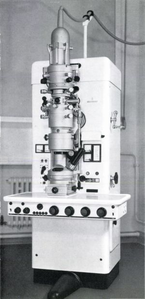
Electron Microscope
General

The Electron Microscope as invented by Ernst Ruska and later refined and diversified by other, is a Microscope which unlike the Light Microscope uses High speed Electrons for the creation of images by various means.
The Electron Microscope can be divided into several Sub Categories
- Transmission Electron Microscope
- Reflection Electron Microscope
- Scanning Electron Microscope
- Emission Electron Microscope
Each of these principles has its own unique advantages and disadvantages. For further information see the related pages.
History
The Electron Microscope was invented in 1934 by German Physascist Ernst Ruska while working for the Siemens Corperation. It evolved from Ruskas findings that a aperture can be magnified by menas of a rotationally symetrical magnetic field, while studying and desining Electron Beam Oscillographs. With the help of Engineer [Max Knoll] and [Bodo von Borries], the observation quickly resulted in a functioning proof of concept aperatus which can be considered the forefather of all electron microscope. All though crude at the time, this apperatus proved the princple discovered by Ruska to be exploitable for the creation of a Microscope.
The apperatus was later Refined numerous times, leading ultimatly to the creation of the [Siemens Elektronen Mikroskop]. This unit was later refined, fallowing World War II into the [ÜM100] Microscope. Simultaniously to the development at Siemens, the German Company AEG with subsequent help from Carl Zeiss, under the leadership of [Otto Rang] and [Hans Schluge], another first generation Microscope was developed. This Microscope subsequently evolved into the AEG Zeiss [EM8]. Unlike the Siemens counterpart, this unit opperated purely by means of the [Electrostatic Lens], where as Ruskas Microscope was based exclusivly on the [Electromagnetic Lens].
Some time latter, the United Kingdom and the American Continent started the development of Electron Microscopes of their own. The British contribution to the world of Electron Microscopy was first started by the Metropolitan-Vickers Corperation, under the leadership of <insert name here>, which created Britains first Electron Microscope, the [EM2].
Meanwhile on the American Continent, the Canadian Scientist James Hillier worked on creating a Microscope of their own, his work was later incorperated into the [RCA] Line of Electron Microscopes.
Some what later the USSR, based on documentation and equipment looted after the conclusion of World War II, begun the development of their own Microscopes. Not much is known about these early Microscopes, but two models have been reported in the <insert name of paper here>. There the <insert first russian microscope here> and <insert Second Microscope here> where discussed. Later Electron Microscope development was moved to what is now the Czchek Republic under the name [Tesla].
Japan also started deveolping Electron Microscopes, two major Companies stood out, JEOL and Hitachi, which still to this day, produce Electron Microscopes of the highest Quality. Hitachi Pioneered the use of the [Cold Field Emmision Cathode], first seen in their <inser name of first FESEM here>.
Types
Transmission Electron Microscope
The Transmission Electron Microscope was the first type of Electron Microscope invented. Its opperating principle is analegous to that of the Compound Light Microscope but (usually) in a invetered fassion, relative to their Light Optical Breatheren. The source of Illumination here, is a Cathode located within the Electron Gun. The resulting stream of Electrons is demagnified and or Re-Focused onto the Sample. The transmitted Eelectrons are then focused by the Objective lens, and the subsequent real images Magnified by the intermediat and or Projector Lens. The final highly magnified image is then projected onto a Scintilating material or Phosphor which converts the Electrons into Photons.
Images may be obtained by either directly immaging the Electrons via Electron Sensetive Film, a second fine grane Screen coupled to a Digital or Analog Camera, or alternativly by means of a Externally mounted Camera focused onto either the Focusing Screen, or Viewing Screen via either the Primary or Secondary Viewport.
Reflection Electron Microscope
The Reflection Electron Microscope is a seldom seen variant of the Electron Microscope, which unlike the Transmission Electron Microscope uses the electrons which the subject under observation reflected in the general direction of the Objective Lens. These electrons are either Inelastically or Elastically Scattered by the Atoms composing the subject.
The general beam path is identical to that of the Transmission Electron Microscope, with the only difference beeing a steep angle between the electron source, Subject and Objetive. Some Transmission Electron Microscopes offer the ability to do Reflection Electron Microscopy all be it at shallow angles, when compared to purpose built machines.
The REM (Reflection Electron Microscope) was the first methode for studiying the native surface of objects at magnifications and Depth of Fields unatanable by classical Light Optical means. The resolution proved to be limmited by the very broud energy spread of the Reflected Electrons. This leads to very a dramatic loss in resolution due to Chromatic Abboration present in all simple Electron Lenses.
Scanning Electron Microscope
The Scanning Electron Microscope was first invented by german Physicist Manfred Baron von Ardenne around 1940. Unlike the two previously discussed types, this Microscope relies not on the focusing of a compleate image onto a View Screen, but rather by means of a sharply focused Electron Probe which is moved accross the Subject.
Various interactions with the Subject can be used for the creation of images, examples include among others, Secondary Electron Imaging, Backscatter Electron Imaging and Transmitted Electrons. Due to the nature of their image formation, their ultimate resolution is somewhat less then that of the Transmission Electron Microscope.
Emission Election Microscope
This Emission Electron Microscope represents a somewhat obscure form of the Electron Microscope. It uses the same basic imaging train as the Transmission Electron Microscope, but rather then bombarding the Subject with Electrons generated in an Electron Gun, the subject its self is made to give off electrons.
The simplest form of the EEM is the experiment of projecting an image of the Electron Source of either a TEM or a Cathode Ray Tube onto a view Screen or Phosphor. Image contrast is formed by the variation Emmision density of the subject.
Samples may be made to release electrions, which must be further accelerated by a ||Electron Gun]] or a Linear Accelerator prior to entering the Objective Lens (if prsent), by means of Heat, Ion Bombardment, Electron Bombardment, or Light Bombardment (such as high energy UV Light).